The Shoulder
Shoulder Girdle
Infiltration of the sternoclavicular joint
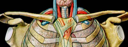
The physician who is planning an infiltration in or nearby the sternoclavicular joint should be completely knowledgeable about some vital structures immediately posterior to the sternoclavicular joint. These vital structures include: the subclavian veins, the innominate artery (right) and the carotis and subclavia arteries (left), the phrenic and vagus nerves.
Technique:
- The patient is in a half-lying position, the shoulders slightly shrugged.
- The medial end of the clavicle and the incisura sterni can easily be papated.
- One palpating finger is brought as deeply as possible behind the joint. This is a safeguard to protect mediastinum and vital structures against a penetrating needle.
- A needle of 2.5 cm is first introduced from in front, penetrating the ligaments at a depth of about 1cm.
- The whole anterior and superior ligaments are infiltrated. Therefore the tip of the needle is partly withdrawn and reinserted several times until 1 cc of the product is injected.
- An infiltration of another one cc of triamcinolone is performed into the posterior sternoclavicular ligament by approaching from above and allowing the needle to progress between the palpating finger and the joint .
Preliminary Examination / Interpretation / Pathology of Shoulder Girdle / Sternoclavicular Lesions
| [Start] | [Main shoulder] | [Flowcharts shoulder] | [A System of Orthopaedic Medicine] |
Copyright © 2021 DR. L. OMBREGT All Rights
Reserved
The author can not be held responsible for any damage caused by the use of any information provided.